Anti Lamin A C Antibody

Rabbit Polyclonal Lamin A C Antibody Stj93885

Cleaved Lamin A Asp230 Antibody Cell Signaling Technology

Anti Lamina C Antibody Rabbit Lamin A C Polyclonal Antibody Np 001244303 1

Anti Lamin A Antibody Ab2559 Abcam

Anti Lamin A Lamin C Antibody Wl4g10 Ko Tested Ab232730 Abcam

Purified Anti Lamin A C Antibody Lamin A C Wl4g10

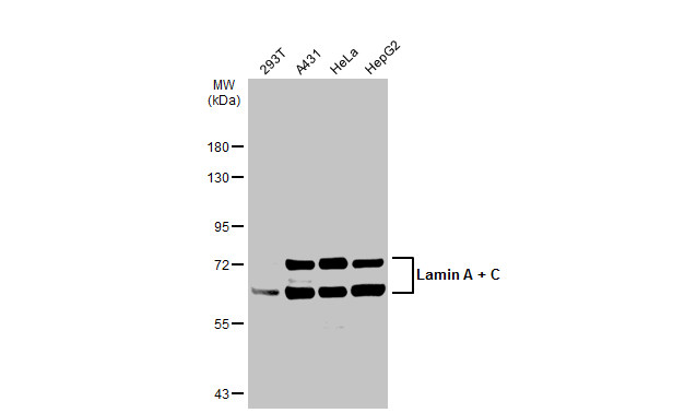
Performed under reducing conditions.
Anti lamin a c antibody.

Purified Anti Lamin A C Antibody Lamin A C Lass2d9

Anti Lamin A Lamin C Antibody Ab224816 Abcam
Anti Lamin A Antibody Ab226198 Abcam
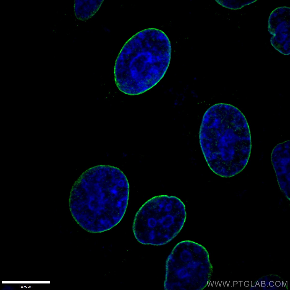
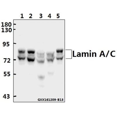
Lamin A C Antibody 10298 1 Ap Proteintech
Anti Lamin A C Antibody Gtx101127 Genetex

Anti Lamin A C Antibody Clone 14 Clone 14 Upstate From Mouse Sigma Aldrich

Anti Prelamin A C Lamin A C Antibody If Validated Bosterbio
Anti Lamin A C Wl4g10 Monoclonal Antibodies Ximbio

Phospho Lamin A C S392 Antibody Ab58528 Abcam
Anti Lamin A C Lass2d9 Antibody 5ml Supernatant Kerafast

Anti Lmna Antibody Mouse Lamin A Cleavage Site Monoclonal Antibody Clone 3a5 Np 733821 1
Anti Lamin A C R386 Antibody A25471 Antibodies Com

Recombinant Anti Lamin A Lamin B1 Lamin C Antibody Epr4068 Ab108922

Anti Progerin Antibody 13a4 Ab66587 Abcam
Lamin A Lamin C Antibody 131c3 Nuclear Envelope Marker Ab8984 Abcam
Lamin B1 Antibody 12987 1 Ap Proteintech

Anti Phospho Lamin A C Ser404 Clone From Rabbit Sigma Aldrich

Ed8fin4g05xqqm

Anti Lamin A C Antibody A12573 Antibodies Com

Anti Lamin A C Antibody Rabbit Lamin A C Monoclonal Antibody P02545
Anti Lamin A C Antibody Center Product No Abin2854943
-Western-Blot-NBP1-50051-img0008.jpg)
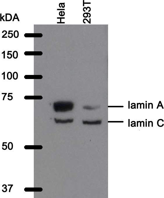
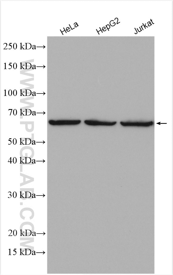
Lamin A C Antibody Em 11 Nbp1 50051 Novus Biologicals

Alexa Fluor 594 Anti Lamin A Lamin C Antibody Epr4100 Ab215324 Abcam
Anti Lamin A C Antibody A80595 Antibodies Com
Source : pinterest.com
